Urgent Care with X-Ray in Port Charlotte, FL
Our goal at PrimeHealth Urgent Care is to meet patients’ expectations by offering quality X-ray services. We can treat the most serious injuries, including complicated fractures and fractured bones. Visit our team for imaging services. We are conveniently located at 23951 Peachland Blvd, Unit 1, Port Charlotte, FL 33954. For more information, contact us or request an appointment online.


Table of Contents:
What to expect when you go for an X-ray?
How do X-rays work step by step?
What should you not do before an X-ray?
How do X-rays see through your body?
When going for an X-ray, the process typically involves checking in at the radiology department and filling out any necessary paperwork. You will probably have to put on a hospital gown and eliminate any jewelry or metal things that could obstruct the imaging. The X-ray technician will position you on a table or stand in a specific way to capture the required images. The technician will then step behind a protective wall and take the X-ray images, which are painless and only take a few seconds. It’s important to remain still during the procedure to ensure clear and accurate images. After the X-rays are taken, they will be reviewed by a radiologist, who will draft a report on the findings. Your healthcare provider will then discuss the findings with you at a follow-up appointment and develop a treatment plan, if necessary, based on the X-ray results.
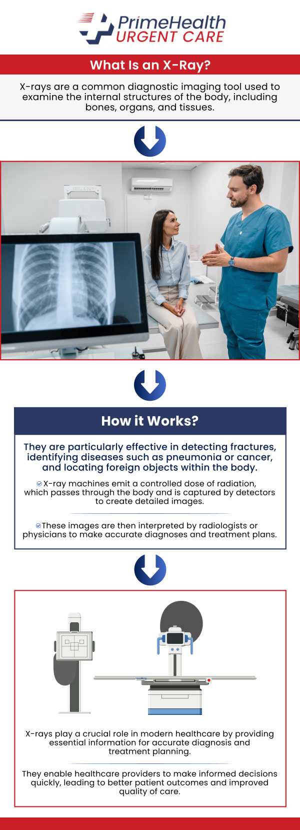
X-rays function by utilizing electromagnetic radiation to create images of the internal structures of the body. The process begins with the X-ray machine emitting a controlled beam of radiation towards the specific body part being examined. This beam of radiation passes through the body, interacting with the various tissues and structures in its path. Dense body parts like bones take in more of the radiation and appear as white areas on the resulting image because they block the radiation from reaching the detector. Conversely, less dense structures, like soft tissues and organs, let radiation pass through more easily and get to the detector, resulting in darker areas on the image. The detector captures the radiation that passes through the body and creates an image that reflects the varying levels of radiation absorption, ultimately producing a detailed visual representation of the internal structures. This image is then reviewed by a radiologist, who interprets it to identify any abnormalities, injuries, or underlying medical conditions. The information obtained from X-ray images helps healthcare professionals make informed diagnoses and develop appropriate treatment plans for patients.
Before undergoing an X-ray procedure, it is essential to take several precautions to ensure the accuracy of the imaging results and maintain the patient’s safety. One important step is to communicate any pertinent medical information to the healthcare provider, such as the possibility of pregnancy, allergies to contrast dye, or any existing health conditions that may affect the imaging process. Additionally, patients should remove any metal objects, such as jewelry, clothing with metal fasteners, or other metallic items, as these can disrupt the X-ray beam and lead to distorted images. Following specific preparation instructions from the healthcare provider is also crucial, such as fasting for a certain period before the X-ray scan or refraining from consuming certain substances that might interfere with the imaging process. By following these suggestions, patients can help with the effectiveness of the X-ray examination, leading to accurate diagnosis and appropriate treatment of any medical issues detected. It is always best to clarify any doubts or concerns regarding the X-ray procedure with the healthcare provider beforehand to promote a smooth and successful imaging experience.
X-rays use high-energy electromagnetic radiation that has a shorter wavelength than visible light, allowing them to penetrate different substances. When directed at the body, X-rays travel through soft tissues relatively unimpeded, as they have lower density and absorb fewer X-rays. However, denser materials like bones and metal absorb more X-rays, resulting in less radiation reaching the detector. This variation in the amount of radiation detected creates an image where bones appear white or light grey due to high absorption, while softer tissues appear darker. X-rays can capture these variations in tissue density and produce detailed images that healthcare professionals can use to identify fractures, tumors, infections, and other abnormalities within the body. Despite their ability to penetrate tissue, X-rays are still harmful in excessive doses, which is why precautions are taken to limit exposure during medical imaging procedures. The development of digital X-ray technology has enhanced image quality, reduced radiation doses, and improved the efficiency of diagnostic processes in healthcare settings, further highlighting the importance of this imaging modality in modern medicine.
Visit PrimeHealth Urgent Care to be examined if you require an X-ray due to any kind of injury, such as a complex fracture or cracked bone. We are conveniently located at 23951 Peachland Blvd, Unit 1, Port Charlotte, FL 33954. For more information, contact us or request an appointment online. We serve patients from Port Charlotte FL, Murdock FL, Harbour Heights FL, Charlotte Harbor FL, and surrounding areas.

Additional Services You May Need

Additional Services You May Need
• Abscesses
• Sports Injuries
• Bug & Animal Bites
• BV Testing
• COVID Testing
• Drug Testing
• EKG Testing
• Flu Shots
• Foreign Body Removal
• Laceration Repair
• Minor Burn Treatment
• Pelvic Exams
• Strep Testing
• STD Testing
• Fractures, Sprains, & Strains
• Splinting Treatment
• School & Sports Physicals
• TB Testing
• Tetanus Shot
• Urgent Care
• Vaccines
• Wound Care
• Work Injuries
• X-Ray
• Internal Lab Services
• Illness/Injury
• Occupational Medicine
• Employment Physicals
• Common Cold
• Auto Injuries
• Weight Loss
• Wegovy
• Mounjaro
• Telehealth
• Stitches
• Respiratory Infection
• Coastguard Physicals
• Sick Visits


